connectivity by design.
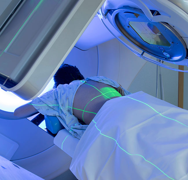
Preventive maintenance is vital in radiotherapy to reduce…
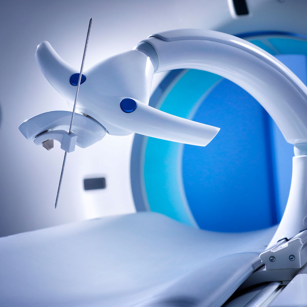
Read MoreLiver cancer is one of the most common and lethal cancers in the world. To diagnose this form of cancer, a physician takes a biopsy by accurately inserting a needle in the tumor. To destroy the tumor, the physician inserts one or more ablation needles. For deep and difficult to reach tumor lesions, this procedure is performed under CT guidance. Even for skilled specialists, it is hard to reach the right spot manually based on the CT image. Sometimes several attempts are necessary to place the needle successfully. To overcome this, a needle placement robot helps the medical specialist find exactly the right angle to place the needle the first time right – to the relief of both patient and specialist.
The starting point for developing the needle placement robot was to retain the current workflow as closely as possible, and only to automate the steps critical for the speed and result (‘first time right’) of the procedure. The critical step is determining the angle at which the needle enters the body.
As a result, we developed a system comprising a head with a needle-guidance mechanism and an arm which secures the head in relation to the operating table with one press of a button. The head can be positioned around the patient manually. Once the system has been positioned around the patient, it accompanies the patient into the CT scanner to determine the position of the patient’s tumor in relation to the head. When determined, the system automatically steers the needle-guidance mechanism to assign it the required direction. The needle is then clamped into the guidance mechanism and inserted into the body by the doctor himself.
Firstly, the system architecture based on the current medical workflow was a challenge. It needed to minimize the barriers to being used by the doctor, both literally and figuratively. A second challenge was the design factor. The entire system, including arm and head, needed to fit in the (tight) space between the patient and the CT scanner’s ring.
However, the biggest hurdle was CT compatibility. The bulk of the system would enter the scanner’s X-ray field but should not disrupt the imaging. That meant that the usual materials like steel, copper and titanium were not an option. We worked with several suppliers including Ceratec (ceramics) and Futura Composites to develop alternatives. Varieties of materials were used in the components, such as composites (for rigid construction parts and flexible elements), ceramics (for highly stressed precision parts and ball bearings), Dyneema fibers (for rotating cable drives), carbon nanotubes (for power wires and switches) and plastic optic fibers (to read out encoder positions remotely).
These solutions imparted high rigidity to the construction, minimizing any transmission play. The result is a system which can guide the needle correctly and precisely, with a margin of error of less than 2 mm at a depth of 25 cm. This means the patient suffers only minimal tissue damage, the medical team can complete the procedure more quickly (and thus more economically), and the doctor remains ‘in control’.

We developed the needle placement system in close collaboration with medical specialists from the Erasmus Medical Center Rotterdam and the University Medical Center Groningen (UMCG). Besides development, we also conducted the clinical study to validate the system. The study included 28 patients at the University Medical Center Groningen (UMCG). The robot-supported method proved to have clear advantages over the manual approach, one prominent advantage being the milder impact on the patient.
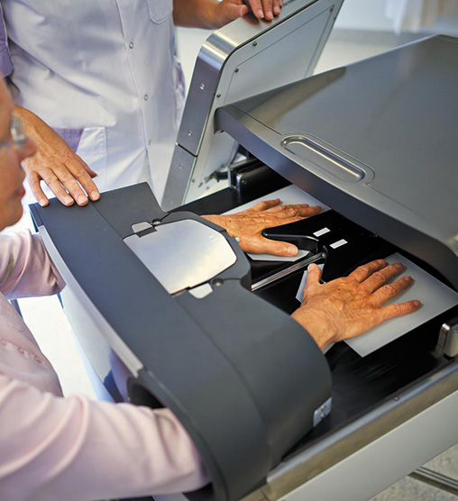
Rheumatoid arthritis is a chronic auto-immune disease, characterized…
Read More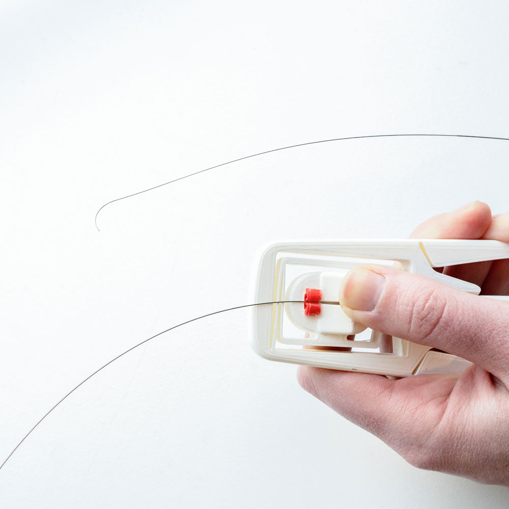
Guidewires have been used for ages to perform…
Read More
Demcon’s task in project MDOT is to develop…
Read More